Persistent pus or blood-stained fluid from an opening near the anus needs interpretation, not simple reassurance. By the end, you will know which drainage patterns need urgent care, what a fistula specialist checks, and how anatomy, continence risk and underlying disease shape the treatment choice.
Key takeaways
- Persistent pus or blood-stained drainage needs examination, not visual self-diagnosis.
- MRI pelvis or examination under anaesthesia can reveal hidden fistula branches.
- Treatment depends on sphincter involvement and the risk to continence.
- Worsening pain, fever, or heavy bleeding after surgery needs urgent medical advice.
What persistent drainage is telling you—and when it is urgent
Ongoing pus or blood-stained drainage usually means an anal fistula tract remains open. A fistula specialist must examine you to confirm the cause; appearance alone cannot establish the diagnosis.
| Pattern | What it may indicate | Clues to discuss |
|---|---|---|
| Open fistula | Persistent drainage | Pus or blood-stained fluid continues through a small skin opening; odour may be unpleasant, with intermittent irritation or swelling. |
| Healing abscess or postoperative wound | Normal recovery | Drainage gradually decreases after drainage or surgery, while pain, redness and swelling also settle. A planned seton or closing cavity can drain for a period. |
| Recurrent abscess | Infection trapped behind a closing opening | Drainage falls after the skin opening closes, then pressure, throbbing pain, swelling or fever develops. Less drainage can signal blockage, not improvement. |
| Infected wound | Wound infection | New or increasing discharge, worsening odour, spreading redness, warmth, tenderness or separation after an operation points to infection. |
| Mucus or stool leakage | Bowel contents reaching the skin | Clear or cloudy mucus can irritate the skin; brown fluid or staining after bowel movements suggests stool leakage. Timing and symptoms still need examination. |
Seek same-day urgent fistula assessment for fever, rapidly increasing pain or swelling, spreading redness, difficulty passing urine, heavy bleeding, or feeling systemically unwell. A blocked external opening can let infection build behind it, so reduced drainage does not prove the fistula is harmless.
How a fistula specialist maps the tract before recommending treatment
A fistula specialist examination combines history, direct inspection, a digital rectal examination and targeted imaging to make a persistent drainage diagnosis. The specialist asks how long drainage has lasted, how much appears, whether it is clear, yellow, bloody or pus-filled, where each skin opening lies, and whether you have had an abscess or operation before.
1. The history also covers bowel pain, diarrhoea, urgency, bleeding, continence, previous sphincter injury or childbirth, and possible Crohn’s disease. Recurrent, multiple or slow-healing fistulas deserve bowel evaluation even when intestinal symptoms are mild.
2. Inspection identifies external openings, scars, redness and active discharge. A digital rectal examination may locate tenderness, an internal opening or a cord-like tract, while sphincter assessment estimates how much muscle the route crosses.
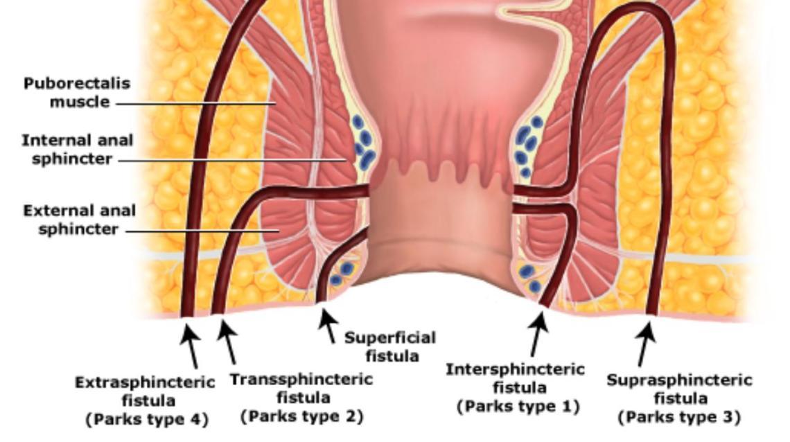
The skin opening does not reveal the full route. An intersphincteric tract stays between the sphincter muscles; a transsphincteric tract crosses the external sphincter; a suprasphincteric tract arches above it; and an extrasphincteric tract lies outside the sphincter complex. A short-looking opening can hide a high route, side branch or horseshoe extension.
Pelvic MRI fistula imaging is especially useful for recurrent, branching, horseshoe, high or Crohn’s-related fistulas. Endoanal ultrasound can clarify the internal opening and sphincter relationship. Examination under anaesthesia helps when pain, scarring or a hidden opening prevents reliable office examination, allowing the surgeon to trace branches before recommending treatment.
How anatomy and continence risk determine the treatment choice
A low, simple tract with little external sphincter involvement often suits fistulotomy: laying the tract open allows it to heal from the base. The shortest procedure is not automatically safest; dividing substantial external sphincter can cause impaired continence, especially when baseline control is already reduced.
| Option | Main purpose | Continence risk and limitation |
|---|---|---|
| Fistulotomy | Opens a carefully selected low tract | Excellent fit for simple anatomy; unsafe when much sphincter would be divided |
| Loose draining seton | Keeps an abscess cavity or tract open to drain and control sepsis | Does not usually eradicate the tract; often a first or staged step |
| Cutting seton | Gradually divides muscle while treating the tract | Different, recognised continence risk because muscle is deliberately divided |
| LIFT, advancement flap or FiLaC | Closes the tract while preserving sphincter | Less predictable healing than a well-selected fistulotomy |
For a tract crossing too much sphincter, sphincter-preserving fistula treatment choices include LIFT, meaning ligation of the intersphincteric fistula tract, an endorectal advancement flap, and laser fistula treatment such as FiLaC where offered and clinically suitable. These avoid deliberate sphincter division, but recurrence remains possible.
Fibrin glue and fistula plugs are non-cutting options with variable durability, not guaranteed cures. Antibiotics treat cellulitis, systemic infection or selected immunocompromised patients; they do not usually close a cryptoglandular tract without source control through drainage and appropriate definitive treatment.
How to interpret drainage after a seton or fistula operation
Persistent drainage after fistula surgery does not by itself prove treatment failure. A healing wound, slowly closing cavity or planned loose seton can continue releasing fluid while inflammation settles.
Track the direction of symptoms rather than judging one day’s amount:
1. Drainage that steadily decreases, with less pain and swelling, fits expected healing. A draining seton may leave a small opening and discharge because it keeps the tract or abscess cavity open.
2. Persistent pus, blood-stained fluid, a continuing external opening, recurrent swelling, worsening pain or a newly visible opening suggests residual, recurrent or inadequately controlled disease. Arrange a review rather than assuming the wound only needs more time.
3. A new painful lump, fever, spreading redness, difficulty passing urine, heavy bleeding or feeling systemically unwell needs urgent assessment. Do not wait for a routine appointment.
A seton often controls sepsis rather than eliminating the tract. It may be a staged step before LIFT, which ligates the intersphincteric fistula tract, an advancement flap or another definitive procedure. A drainage reduction followed by swelling can mean the skin opening has closed while infection is trapped behind it.
Repeated operations can fail when Crohn’s disease drives ongoing inflammation. Recurrent, multiple, complex or unusually slow-healing fistulas deserve bowel evaluation; a Crohn’s disease fistula may need biologic therapy alongside drainage and surgery. Any recurrent fistula treatment plan should address the underlying inflammation, not repeat operations alone.
What to ask before choosing a fistula treatment plan in Mumbai
Compare a proposed plan on three points: chance of durable healing, recovery demands and risk to bowel control. The shortest recovery is not automatically the safest choice.
| Option | Main trade-off | Questions to ask |
|---|---|---|
| Fistulotomy | Strong option for a low, simple tract; divides involved muscle | Is enough external sphincter spared? |
| LIFT or advancement flap | Preserves sphincter; healing is less predictable | Why does this anatomy favour it? |
| FiLaC, fibrin glue or plug | Avoids deliberate muscle division; durability varies | What is the plan if the tract remains open? |
| Loose seton | Controls drainage and sepsis; may precede definitive treatment | Is it temporary, or intended to remain longer? |
Take this checklist to your fistula treatment consultation:
- Where is the internal opening, and how much external sphincter does the tract cross?
- Is there a side branch, horseshoe extension or hidden abscess cavity?
- Do I need pelvic MRI, endoanal ultrasound or examination under anaesthesia?
- Why is fistulotomy, LIFT, an advancement flap, FiLaC or another option suitable for this anatomy?
- How do my baseline continence, childbirth or prior sphincter injury, smoking, diabetes, previous operations and possible Crohn’s disease change the recommendation?
A fistula specialist Mumbai patients consults should balance durable healing against continence risk, not promise a guaranteed minimally invasive cure. Dr. Jiten Chowdhry can provide specialist assessment for persistent drainage and discuss suitable sphincter-preserving options, but examination and, when needed, imaging determine the plan.
Related items
Fistula Treatment An fistula is a small abnormal tunnel that develops between the inside of the anal canal and the skin around the anus. View service → |
 | Anal Fistula When an irregular false tract occurs between the inside of the rectum and the outside skin surface, an Anal Fistula forms. View product → |
Frequently asked questions
When does persistent fistula drainage require urgent medical attention?
Seek urgent advice for fever, worsening pain or swelling, heavy bleeding, difficulty passing urine, or feeling faint. Ongoing pus or blood-stained drainage also needs examination because the tract may remain open.
How does a fistula specialist map the tract before treatment?
The specialist combines an examination with selected tests, such as MRI of the pelvis or examination under anaesthesia, to locate the internal opening, side branches, abscesses, and sphincter involvement.
How does anatomy affect the choice of fistula treatment?
The treatment depends on how much anal sphincter muscle the tract crosses, whether branches or an abscess exist, and your baseline continence. Cutting through more muscle can increase continence risk.
What drainage is normal after a seton or fistula operation?
Some drainage can continue while a seton remains or during wound healing. Contact your surgeon if drainage increases with fever, escalating pain, new swelling, heavy bleeding, or a sudden change in the wound.