The external opening does not reveal the full fistula tract or show how much anal sphincter muscle it crosses. You will understand how a fistula specialist examines and maps the tract, what happens during laser closure, how recovery is monitored, and when another treatment is safer.
Key takeaways
- Ask how much anal sphincter the fistula crosses before choosing treatment.
- Confirm whether MRI pelvis or endoanal ultrasound is needed.
- Expect tract cleaning, laser closure and follow-up after surgery.
- Seek urgent review for fever, worsening pain, swelling or pus.
How the first consultation confirms what needs treatment
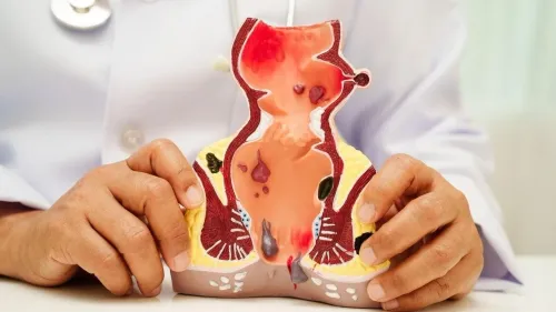
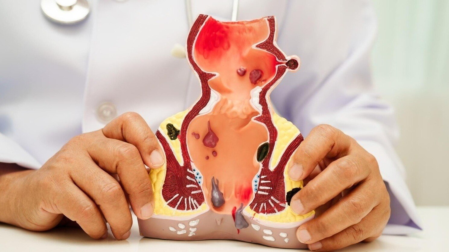
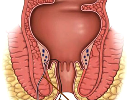
A fistula specialist first confirms the diagnosis, traces the likely route, and assesses how much anal sphincter the tract crosses before recommending laser closure. The external opening alone cannot show whether the fistula is low, branching, recurrent, or linked to an abscess.
The first examination usually covers:
- Your history: drainage, pain, swelling, fever, previous abscess drainage or fistula surgery, bowel disease such as Crohn’s disease, and any change in continence.
- External inspection: the number and position of skin openings, redness, discharge, scars, induration, and a tender lump suggesting an abscess.
- Digital rectal examination: tenderness, a cord-like tract, the internal opening, sphincter tone, and any mass inside the anal canal.
- Anoscopy or proctoscopy when appropriate: direct inspection of the anal canal to look for the internal opening and other disease.
- Examination under anaesthesia if pain, scarring, or complex anatomy prevents a reliable office examination.
An abscess changes the immediate plan: infected fluid usually needs drainage and sepsis control before elective closure. A loose draining seton may be used to keep the tract open and control drainage; it is not usually the definitive cure.
Before consent, ask which tract classification applies, how much sphincter is at risk, and why laser is preferable to fistulotomy or another sphincter-sparing method. The specialist should also explain follow-up and what happens if the tract remains open.
How imaging and examination map the fistula before surgery
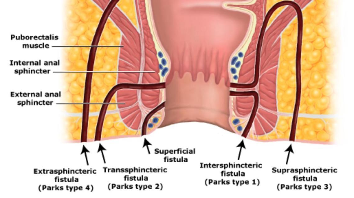
The plan begins with anatomy, not the external opening alone. A fistula specialist inspects the skin, performs a digital rectal examination and uses anoscopy or proctoscopy to look for pus, tenderness and the suspected internal opening. The examination also indicates whether the track may pass through the sphincter.
| Test | What it can show | When it helps |
|---|---|---|
| MRI pelvis with fistula protocol | The main tract, internal opening, side branches, abscesses, horseshoe extensions and the tract’s position relative to the sphincter | Complex, recurrent, high or branching fistulas |
| Endoanal ultrasound | The internal opening and the layers of the internal and external sphincter crossed by the tract | When sphincter detail is needed or MRI is unsuitable |
| Examination under anaesthesia | The openings, tract direction and tissue planes through gentle probing, dye or injection | When pain, scarring or uncertainty prevents a reliable clinic examination |
| Anoscopy or proctoscopy | The anal canal and a possible internal opening | During the clinical assessment when the opening is close enough to see |
No single scan decides whether laser closure is suitable. The specialist combines the examination, imaging and continence history to estimate how much sphincter is at risk. If the internal opening remains uncertain, examination under anaesthesia can prevent laser treatment aimed at the wrong tract.
What happens during laser fistula closure in the operating room
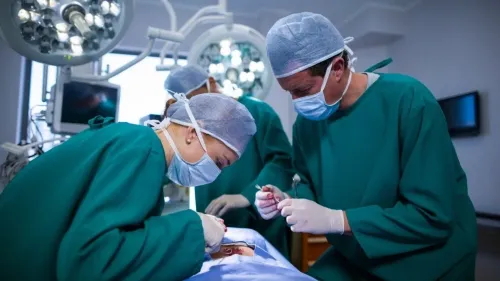
During FiLaC, the surgeon passes a radial-emitting laser fibre through the external opening toward the internal opening, then withdraws it while delivering energy. The heat contracts and seals the tract from within. The internal opening is usually closed or treated separately; the laser does not simply erase the tract from the skin outward.
If an abscess or uncontrolled infection is present, drainage takes priority. A fistula specialist may place a loose seton before definitive closure to keep pus draining and control sepsis.
| Option | What happens | Main trade-off |
|---|---|---|
| FiLaC | Laser energy closes the tract while preserving the sphincter | The tract can remain open or recur, requiring follow-up or another procedure |
| Fistulotomy | The surgeon lays the tract open so it heals as a groove | Highly effective for selected low fistulas, but dividing substantial sphincter can threaten continence |
| Seton placement | A thread maintains drainage through the tract | Usually controls infection rather than providing definitive closure |
| LIFT | The surgeon reaches the tract between sphincter muscles, ties it off and divides it | Sphincter-sparing, but unsuitable for every branch or internal opening |
| Advancement flap | Tissue is lifted and moved over the internal opening after tract treatment | Preserves sphincter, but healing can fail and the operation is more extensive |
The choice depends on sphincter involvement, internal-opening position, branches, horseshoe extensions, Crohn’s disease, previous operations, recurrence and continence. Your specialist should explain why FiLaC suits that anatomy, what follow-up will check, and what happens if the opening remains.
What recovery, follow-up and warning signs look like
Soreness, light bleeding and discharge can occur after FiLaC, particularly while the external opening heals. Keep the area clean, follow the wound-care instructions, take prescribed pain relief, and prevent hard stools with fluids, fibre or a laxative recommended by your clinician. Ask when you can return to work, exercise and sexual activity.
Follow-up checks confirm whether healing is progressing rather than assuming that a closed skin opening means success. A fistula specialist will ask about pain and drainage, inspect the external opening, and assess tenderness, swelling or a palpable tract. Digital rectal examination and anoscopy can help assess the internal opening when appropriate.
Persistent symptoms or uncertain findings may require a pelvic MRI with fistula protocol or examination under anaesthesia.
Contact your surgical team promptly if you develop:
- Fever or chills
- Increasing pain or a new painful swelling
- Pus, foul-smelling drainage or heavy bleeding
- An external opening that stays open or repeatedly discharges
- Difficulty passing urine or stool
These findings can indicate an abscess, a fistula that remains open, or recurrence. The specialist may first drain an abscess, then use imaging to locate residual or branching disease.
If the tract has not closed, the plan could include continued drainage, a seton or another sphincter-sparing procedure; repeat treatment is part of fistula care, not proof that the original diagnosis was wrong.
When laser is a sensible choice—and what to ask before consenting
Laser is sensible when the tract can be cleaned and closed without leaving uncontrolled infection or sacrificing important sphincter muscle. A fistula specialist bases that decision on the internal opening, the amount of sphincter crossed, branching or horseshoe extensions, Crohn’s disease, previous operations, recurrence and your continence status—not on the external opening alone.
A low, straight tract with little sphincter involvement is often better treated by fistulotomy, which opens the tract to heal from the inside. A high or complex tract may need laser closure, LIFT, an advancement flap or staged treatment. If an abscess or heavy infection is present, drainage comes first.
A loose seton may control sepsis while the tissues settle; it drains the fistula but is not usually the final cure.
Before consenting, ask:
- What is the exact fistula classification, and where is the internal opening?
- How much external and internal sphincter does the tract cross?
- Do MRI pelvis, endoanal ultrasound or examination under anaesthesia change the plan?
- Why is laser preferable to fistulotomy, LIFT, a flap or continued seton drainage in my case?
- Is branching disease, Crohn’s disease or previous surgery likely to reduce healing?
- What will you do if the tract remains open or returns, and how long will follow-up continue?
Whether you consult Dr Jiten Chowdhry or another surgeon, request a clear plan for drainage, continence protection and failure—not just claims about quick recovery or minimal scarring.
Related products
 | Laser Surgery For Fistula We are best Fistula Treatment Surgery Doctor In Mumbai . Get Guidance from Medical experts to select laser surgery for fistula in Mumbai from trusted... View product → |
 | Anal Fistula When an irregular false tract occurs between the inside of the rectum and the outside skin surface, an Anal Fistula forms. View product → |
Frequently asked questions
How does a fistula specialist confirm what needs treatment?
The specialist reviews your symptoms, examines the external and internal openings, checks for abscess, and assesses whether the tract is low, branching or recurrent. The external opening alone cannot define the full fistula.
How is a fistula mapped before laser surgery?
The specialist combines examination with imaging when the route is unclear, the fistula is recurrent or branching, or an abscess is suspected. MRI pelvis and endoanal ultrasound can show the tract and its relationship to the anal sphincter.
What happens during laser fistula closure?
After anaesthesia, the surgeon identifies and cleans the tract, treats any associated abscess, and uses a laser fibre to close the tract from inside. The exact steps depend on the fistula’s anatomy.
What warning signs need medical review after surgery?
Contact your surgical team for fever, increasing pain or swelling, heavy bleeding, difficulty passing urine, worsening discharge or a new lump near the anus.
When is laser fistula closure a sensible choice?
Laser closure is worth discussing when the fistula’s anatomy is suitable and preserving anal sphincter function is a priority. Ask about the expected success rate for your tract, alternatives, recurrence risk and the plan if closure fails.
Related items
 | Laser Treatment Hemorrhoids /Piles Hemorrhoids : Hemorrhoids are varicose (enlarged and swollen) veins around the outside of the anus or in the lower rectum. View product → |
 | When the skin of the anus gets cut or damaged, that is called an anal fissure. It is very similar to what happens when our dry lips get chaffed. View product → |
Hernia Treatment What is a Hernia? A hernia develops when a portion of the intestine or abdominal tissue protrudes through a weakened abdominal wall. View service → |
 | VARICOSE VEINS TREATMENT WITH ENDOVENOUS LASER ABLATION What does it mean varicose veins endovenous ablation? vein-ablation-procedureVaricose pathology and its best methods of treatment have been developed... View product → |